Biological Sciences
Gram Stain
Gram stain is a laboratory technique used to differentiate bacteria into two groups based on their cell wall composition. It involves staining bacterial cells with crystal violet, iodine, alcohol, and safranin. Gram-positive bacteria retain the purple color, while gram-negative bacteria appear pink after the staining process. This method is widely used in microbiology to quickly identify and classify bacteria.
Written by Perlego with AI-assistance
Related key terms
1 of 5
10 Key excerpts on "Gram Stain"
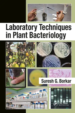 eBook - ePub
- Suresh G. Borkar(Author)
- 2017(Publication Date)
- CRC Press(Publisher)
The Gram Stain is a differential stain developed by Dr. Hans Christian Gram, a Danish physician, in 1884. The staining is called Gram Staining, after Dr. Gram. It is a very useful stain for identifying and classifying bacteria into two major groups: Gram-positive and Gram-negative.In this process, the fixed bacterial smear is subjected to four different reagents in the order listed: Crystal violet (primary stain), iodine solution (mordant), alcohol (decolorizing agent), and safranin (counterstain). The bacteria which retain the primary stain (appears dark blue or violet) are called Gram-positive, whereas those that lose the crystal violet and get counterstained safranin are referred to as Gram-negative.The differences in staining responses to the Gram Stain can be related to the chemical and physical differences in the bacterial cell walls. The Gram-negative bacterial cell wall is a thin, complex, multilayered structure and contains relatively high lipid content, in addition to protein and mucopeptides. The higher amount of lipids is readily dissolved by alcohol, resulting in the formation of large pores in the cell wall that do not close appreciably on dehydration of cell-wall proteins, thus facilitating the leakage of the crystal violet–iodine (CV-I) complex and resulting in the decolorization of the bacterium, which later takes the counterstain and appears red. In contrast the Gram-positive cell walls are thick and chemically simple, composed mainly of protein and cross-linked mucopeptides. When treated with alcohol, it causes dehydration and closure of cell wall pores, thereby not allowing the loss of the CV-I complex, and so the cells remain purple.Material RequiredCultures of test bacterium that are 24 or fewer hours old, Gram Staining reagents crystal violet, Gram’s iodine solution, 95 percent ethyl alcohol, safranin, staining tray, wash bottle of distilled water, droppers, inoculating loop, glass slides, blotting paper/absorbent paper, tissue paper, Bunsen burner/spirit lamp, microscope.ProcedureMake thin smears of test bacterium on glass slides. Let the smears air dry. Heat fix the smears. Hold the smears using a slide rack or clothes pin. Cover the smear with crystal violet for 30 seconds. Wash the slide with distilled water for a few seconds using the wash bottle. Cover the smear with Gram’s iodine solution for 60 seconds. Wash off the iodine solution with 95 percent ethyl alcohol. Add ethyl alcohol drop by drop, until no more color flows from the smear. (The Gram-positive bacteria are not affected while all Gram-negative bacteria are completely decolorized.) Wash the slides with distilled water and drain. Apply safranin to smears for 30 seconds (counterstaining). Wash with distilled water and blot dry with absorbent paper. Let the stained slides air dry. eBook - PDF
- Osei, Getachew(Authors)
- 2018(Publication Date)
- Agri Horti Press(Publisher)
This ebook is exclusively for this university only. Cannot be resold/distributed. 17 Microscopy: An Introduction An example is the Gram Stain technique. This differential technique separates bacteria into two groups, Gram-positive bacteria and Gram-negative bacteria. Crystal violet is first applied, followed by the mordant iodine, which fixes the stain. Then the slide is washed with alcohol, and the Gram-positive bacteria retain the crystal-violet iodine stain; however, the Gram-negative bacteria lose the stain. The Gram-negative bacteria subsequently stain with the safranin dye, the counterstain, used next. These bacteria appear red under the oil-immersion lens, while Gram-positive bacteria appear blue or purple, reflecting the crystal violet retained during the washing step. Fig . The Gram Stain Procedure used for Differentiating Bacteria into Two Groups. Another differential stain technique is the acid-fast technique. This technique differentiates species of Mycobacterium from other bacteria. Heat or a lipid solvent is used to carry the first stain, carbolfuchsin, into the cells. Then the cells are washed with a dilute acid-alcohol solution. Mycobacterium species resist the effect of the acid-alcohol and retain the carbolfuchsin stain. Other bacteria lose the stain and take on the subsequent methylene blue stain. Thus, the acid-fast bacteria appear bright red, while the non-acid-fast bacteria appear blue when observed under oil-immersion microscopy. Other stain techniques seek to identify various bacterial structures of importance. For instance, a special stain technique highlights the flagella of bacteria by coating the flagella with dyes or metals to increase their width.Flagella so stained can then be observed. A special stain technique is used to examine bacterial spores. Malachite green is used with heat to force the stain into the cells and give them colour. A counterstain, safranin, is then used to give colour to the non-sporeforming bacteria.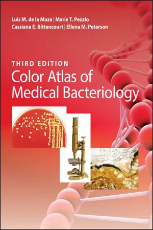 eBook - ePub
- Luis M. de la Maza, Marie T. Pezzlo, Cassiana E. Bittencourt, Ellena M. Peterson(Authors)
- 2020(Publication Date)
- ASM Press(Publisher)
41Stains, Media, Reagents, and HistopathologyBacteria are identified in the clinical laboratory by a variety of methods, including microscopy, observation of growth characteristics, determining reactions to organic and inorganic compounds, and molecular techniques. Examination of histopathological sections can also contribute to the definitive diagnosis or exclusion of bacterial infections. This chapter presents images of the more common stains, media, and tests used in the diagnostic medical microbiology laboratory, as well as relevant histopathologic images. Gram Staining is the most widely used method for visualizing bacteria. Shown in this chapter are examples of the various sizes, shapes, and arrangements of both aerobic and anaerobic bacteria found in clinical specimens. Also included are some of the most common culture media used in the laboratory for demonstrating growth, isolation, colonial morphology, hemolysis, pigment production, and other distinguishing bacterial characteristics. The figures depict some of the key tests and histologic images that have been found to be most helpful for the detection and identification of clinical isolates.STAINS
Gram Stain
Gram Staining is one of the most important procedures in bacteriology. It is used to classify bacteria based upon their size, shape, arrangement, and Gram reaction. It was originally described in 1884 by Christian Gram; the modification currently in use was developed by G. J. Hucker in 1921. The reagents and stains used in Hucker’s modification include crystal violet, Gram’s iodine, acetone alcohol, and safranin. The figures in this section demonstrate a variety of morphotypes as well as Gram Stain reactions.Hematoxylin and Eosin Stain
Hematoxylin and eosin (H&E) stain is used routinely in histopathological sections, as it displays the tissue morphology. Hematoxylin dyes the host cell nucleus with a deep‐purple blue color, and eosin stains the host cytoplasm and the extracellular matrix with a variable degree of pink color. Microorganisms, in particular bacteria and fungi, are also stained with H&E.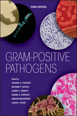 eBook - ePub
- Vincent A. Fischetti, Richard P. Novick, Joseph J. Ferretti, Daniel A. Portnoy, Mirian Braunstein, Julian I. Rood, Vincent A. Fischetti, Richard P. Novick, Joseph J. Ferretti, Daniel A. Portnoy, Mirian Braunstein, Julian I. Rood(Authors)
- 2019(Publication Date)
- ASM Press(Publisher)
The Gram-Positive Bacterial Cell Wall
Manfred Rohde 1HISTORICAL BACKGROUND
In 1884, the Danish bacteriologist Hans Christian Gram developed a staining procedure to view stained bacteriaunder the light microscope (1 ). His staining method, nowadays simply called Gram Staining, discriminated between a Gram-positive and Gram-negative bacterial cell wall. He introduced a dye, gentian violet, which penetrates the cell wall and cytoplasmic membrane, thus staining the cytoplasm of the heat-fixed bacteria. After addition of iodine, an insoluble complex is formed which is retained by the Gram-positive bacterial cell wall upon addition of a decolorizer such as ethanol. Therefore, Gram-positive bacteria appear almost purple, while Gram-negative bacteria retain the dye to a lesser extent or not at all and have to be counterstained with a second dye, safranin or fuchsine, appearing pink or reddish. It is noteworthy that some mycobacteria showed an indifferent staining behavior when Gram Stained, suggesting that the cell wall of mycobacteria might be somehow different from the other two types. In the following decades, it became obvious that cell walls/cell envelopes are more diverse and that Gram Staining alone often could lead to misinterpretations of the cell wall composition.Before the early 1950s, when the chemical composition of bacterial cell walls was not known, it was speculated that chitin or cellulose, polymers recognized as providing rigid structures to other organisms, might also represent the building material of the bacterial cell wall. In 1951, experiments with a phenol-insoluble material from Corynebacterium diphtheria (2 ) revealed glucosamine and diaminopimelic acid as components of the bacterial cell wall which are associated with polysaccharides. Chemical examination of streptococcal cell wall layers highlighted the presence of amino acids and hexosamines in the cell wall extract, as well as rhamnose as a main component in Gram-positive bacteria (3 , 4 ). Systematic analyses of a number of Gram-positive bacteria identified the hexosamines glucosamine and muramic acid as major components together with three prevalent amino acids, namely, d-alanine, lysine or diaminopimelic acid, and glutamic acid. By then, a typical basic basal unit in Gram-positive cell walls was also recognized in which glucosamine and muramic acid are linked with three amino acids via a peptide bond (5 , 6 ). Gram-negative bacteria express the identical basal unit. Numerous analyses of other bacteria revealed that each bacterial genus or even species is often characterized by a distinctive pattern of amino acids, amino sugars, and sugars connected to the basic basal unit. It was believed that these differences should provide a valuable pattern to discriminate between bacterial genera/species (7 , 8 ). Over the following years other compounds of the Gram-positive cell wall were recognized, such as teichoic acids (TAs), which are polyribitol phosphates (9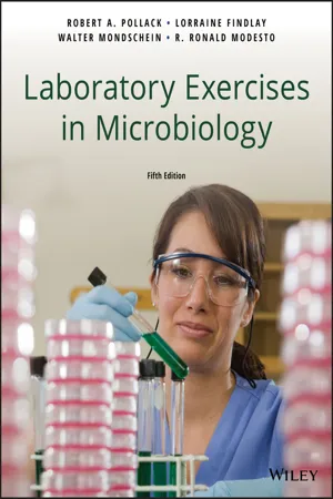 eBook - PDF
- Robert A. Pollack, Lorraine Findlay, Walter Mondschein, R. Ronald Modesto(Authors)
- 2018(Publication Date)
- Wiley(Publisher)
When observed under the microscope, the cells exhibit a blue color. This indicates that these cells are: a. Gram-positive b. Gram-negative c. acid-fast d. nonacid-fast You were introduced to the simple stain in Exercise 3, where all the microbes seen with the microscope were the color of the single stain used for the preparation. Differential staining requires more than one type of stain and is used to distinguish between various types of bacterial cells. A differential stain typically consists of three main steps: first, a primary stain, which is used to stain all the cells on the slide; then a decolorizing step, which removes the stain only from certain types of cells; and finally, a counter- stain, which stains the newly decolorized cells but has no effect on the cells still holding the primary stain. The Gram Stain and acid-fast stain are two widely used differential stains used to distinguish and classify bacteria according to their cell walls. THE Gram Stain Hans Christian Gram (1853–1938) discovered and per- fected the Gram Stain in the 1880s while working on a technique to detect mammalian cells infected with bacteria. 54 EXERCISE 5 Gram Stain and Acid-Fast Stain His discovery was soon used to divide bacteria into two main groups––Gram-positive and Gram-negative— as well as two smaller groups—Gram-nonreactive and Gram-variable. The Gram Stain reaction is based on the amount of peptidoglycan found in the cell walls of these bacteria. Gram-positive bacteria have many layers of pep- tidoglycan, which, in turn, holds molecules of teichoic acids. Gram-negative bacteria have only one layer of pep- tidoglycan with no teichoic acid. Teichoic acid reacts with the crystal violet and iodine used in this staining process. A complex of crystal violet–iodine–teichoic acid mole- cules forms, which results in large, difficult-to-remove complexes. eBook - ePub
Microbial Food Safety
A Food Systems Approach
- Charlene Wolf-Hall, William Nganje(Authors)
- 2017(Publication Date)
- CAB International(Publisher)
6 Gram-Positive BacteriaKey Questions• Which Gram-positive bacteria are of most concern for microbial food safety?• What are the mechanisms by which these Gram-positive bacteria cause illness?• What are the hazards these Gram-positive bacteria present to consumers?• What controls are available to prevent foodborne illness due to these Gram-positive bacteria?The Difference Between Gram-Positive and Gram-Negative Bacteria
Hans Christian Gram was the Danish scientist who, in 1884, published a method for a staining technique to help better see bacteria in tissue samples under the microscope. An unanticipated result of this technique was a way to differentiate two major groups of bacteria based on their cell wall compositions.Bacterial cell membranes that contain thick layers of peptidoglycan are able to retain the crystal violet stain used in the method, resulting in purple- or violet-stained cells that are described as Gram positive. Bacteria that contain less peptidoglycan in their cell membranes are unable to retain the crystal violet stain after the destaining step of the procedure, and as a result of counter-staining with safranin dye appear red or pink, and are described as Gram negative. As with all microbiological testing methods, there are limitations and some bacterial species may produce Gram-variable results, indicating an ability to stain with either reaction result and not provide a clear distinctive result.Gram Staining is a preliminary test used on bacterial cultures to give clues to the identity of the species. The Gram reaction is a first clue, and then other clues like the microscopic cell morphology or shape and placement can provide other clues. The remainder of this chapter will focus on those bacterial pathogens of most concern in foods that fall under the category of Gram positive, and Chapter 7 eBook - ePub
Handbook of Textile and Industrial Dyeing
Volume 2: Applications of Dyes
- M Clark(Author)
- 2011(Publication Date)
- Woodhead Publishing(Publisher)
Colouring biological samples is similar, in scientific principle, to textile dyeing. In both areas, dyes are added to a material with the object of dye molecule attachment, usually to a polymeric substrate. However, where in textiles the chemical make-up of the substrate is well established – cotton/cellulose, silk/protein, etc. – allowing considerable control of the resulting colour and shade, biological samples are usually a complex chemical mixture, the resulting colour and colour distribution depending on the relative proportions of different biopolymers. The proportion and distribution of these biopolymers will also vary depending on the growth phase and age of the cell.As mentioned above, the Gram Staining procedure provides the cornerstone of rapid identification of bacteria in our hospitals. The process was developed by the Dane, Christian Gram in 1884 – pioneering work in an era which was less than accepting of the ‘Germ Theory’ of disease. Such research, built upon by contemporary scientists such as Robert Koch, Paul Ehrlich and others (Leishmann, Romanovsky, Giemsa – the list is fairly substantial) eventually developed into the scientific branch of histology (Wainwright, 2003b ). Specialisms within histology include histopathology, i.e. the study of disease processes via staining, which is really what Gram and subsequent workers set out to do. Modern medical laboratory scientific officers carry out such bacterial staining routinely, alongside further biochemical testing to give information on specific strains of bacteria, particularly in terms of drug susceptibility.Biological stains thus remain of immense importance in modern medicine from the point of view of infection control. However, cell staining is much more extensive than just the differentiation of bacterial and human cells.Usually, medical microbiologists examine cell samples taken from patients. These samples normally include blood, sputum, urine, faeces or tissue swabs, depending on the disease presentation. The process then runs along the lines of ‘Is there an infective organism here? If so, what is it?’ Other disease states may not be so easy to pinpoint – for example where the problem lies in human cell differentiation. Obviously, if such differentiation is required within the live patient, there are more stringent requirements in terms of dye toxicity (q .v eBook - ePub
- Stephen P. Denyer, Norman A. Hodges, Sean P. Gorman, Brendan F. Gilmore, Stephen P. Denyer, Norman A. Hodges, Sean P. Gorman, Brendan F. Gilmore(Authors)
- 2011(Publication Date)
- Wiley-Blackwell(Publisher)
Observation of stained and wet preparations of clinical specimens (blood, pus, sputum) and isolated pure cultures of bacteria from the manufacturing environment provides rapid and essential information to guide further identification. The application of simple stains such as the Gram Stain can divide the various genera of bacteria into two convenient broad groups. The size and shapes of individual cells and their arrangement into clusters, chains and tetrads will also guide identification, as will specific stains for the presence of endospores, capsules, flagella and inclusion bodies. Examination of wet preparations can give an indication as to the motility status of the isolate, and these procedures all represent an important first stage in the identification process.8.3 Biochemical testing and rapid identificationThe differing ability of bacteria to ferment sugars, glycosides and polyhydric alcohols is widely used to differentiate the Enterobacteriaceae and in diagnostic bacteriology generally. Fermentation can be indicated by pH changes in the medium with or without gas production visualized by the collection of bubbles in inverted tubes. More specialized media examine the ability of certain strains to oxidize or reduce particular substrates. There are many hundreds of individual biochemical tests available that each separately seek the presence of a particular enzyme or physiological activity. Taxonomic studies have led to the recognition that certain of these tests in combination characterize particular species of bacteria. Various manuals such asBergey’ s Manual (Holt, 1994) and Cowan and Steel’s Manual for the Identification of Medically Important Bacteria (Barrow & Feltham, 2004) provide a logical and sequential framework for the conduct of such tests. Identification of particular species and genera by such processes is time-consuming, expensive and may require numerous media and reagents.This process has become simplified in recent years by the development of rapid identification methods and kits. The latter often use multiwell microtitration plates that can be inoculated in a single operation either with an inoculated wire or with a suspension of a pure culture. Each individual well contains the medium and reagents for the conduct of a single biochemical test. Identification kits vary in their complexity and also in the precision of the identification made. Simple kits may perform only 8–15 tests, more complex ones are capable of performing 96 simultaneous biochemical evaluations. Scoring of each test and entry into a computer database then allows the pattern of test results to be compared with a large panel of organisms and a probability of identity calculated. As different sets of tests will be required for different classes of bacteria, guidance as to the initial choice of kit is given on the basis of the Gram Stain reaction, and the results of oxidase and catalase tests performed directly on isolated colonies. In large diagnostic laboratories and in quality assurance laboratories automated systems are deployed that can inoculate, incubate and analyse hundreds of individual samples at a time. eBook - PDF
- James E. Moore Jr, Duncan J. Maitland(Authors)
- 2013(Publication Date)
- CRC Press(Publisher)
exploits the difference between types of bacteria in the solubility of trapped dye molecules� Gram-positive bacteria retain the dye after treatment with iodine and appear blue, whereas Gram-negative organisms appear colorless and may then be visualized by treatment with a counterstain, usually neutral red, to the background� The pres-ence or absence of this stain together with the shape of the bacterium provides the pathologist with a simple method of classifying microorganisms found in infectious diseases and infected wounds� FIGURE 13.7 ( See color insert. ) Section through the lining of the small intestine stained with hematoxylin and eosin� eBook - ePub
Microbiological Examination Methods of Food and Water
A Laboratory Manual, 2nd Edition
- Neusely da Silva, Marta H. Taniwaki, Valéria C.A. Junqueira, Neliane Silveira, Margarete Midori Okazaki, Renato Abeilar Romeiro Gomes(Authors)
- 2018(Publication Date)
- CRC Press(Publisher)
Prepare a smear of the culture. If the culture is in a solid medium, place a drop of a saline solution (NaCl 0.85%) on a clean and dry glass slide and emulsify a loopful of the culture with the saline solution. If the culture is contained in broth, agitate the broth and place a loopful on the glass slide. Wait until the liquid dries naturally and then heat-fix the smear by passing the slide through the flame of a Bunsen burner three times. Complete the staining procedure the following way: cover the smear with Hucker’s crystal violet solution and maintain the contact between the smear and the solution for 1 minute. Wash in running water and cover with iodine solution (Lugol) for 1 minute. Wash in running water and then flush with ethanol 95% until no colorant runs off (30 seconds). Wash in running water and cover with safranin solution for 10 seconds. Wash in running water and dry. Observe under an optical microscope using an oil immersion objective. Gram-positive bacterial cells will become purple colored and the Gram-negative cells will turn red. In both cases, the morphological characteristics of the cells as well as their arrangement and appearance can be observed.5.3.5.2 KOH test for confirmation of doubtful Gram Staining (Gregersen, 1978)In some situations the result of Gram Staining is doubtful. In these cases the KOH test, also called the string test, can be used to confirm the result. Place one to two drops of 3% potassium hydroxide (KOH) solution on a clean, dry glass slide. Transfer a loopful of the culture to the drop of KOH and emulsify well. Examine within 60 seconds for cell lyse (loss of turbidity), which allows the DNA from Gram negative bacteria to string when a loopful is lifted (up to 2 to 3 cm) from the slide (KOH positive test).- Note e.2.1) Some species of Gram-positive bacteria may give doubtful or erroneous results, appearing as KOH positive, such as strains of Bacillus macerans (Gregersen, 1978), B. cereus (Car-lone et al ., 1983), B. megaterium and Thermoanaerobacter (Wiegel and Quandt, 1982). Similarly, some Gram-negative bacteria appear KOH negative, such as Xanthobacter strains (probably due to capsule presence) (Wiegel and Quandt, 1982) and Moraxella strains (Carlone et al
Index pages curate the most relevant extracts from our library of academic textbooks. They’ve been created using an in-house natural language model (NLM), each adding context and meaning to key research topics.