eBook - ePub
Atlas of Oral and Maxillofacial Radiology
Bernard Koong
This is a test
- English
- ePUB (handyfreundlich)
- Über iOS und Android verfügbar
eBook - ePub
Atlas of Oral and Maxillofacial Radiology
Bernard Koong
Angaben zum Buch
Buchvorschau
Inhaltsverzeichnis
Quellenangaben
Über dieses Buch
The Atlas of Oral and Maxillofacial Radiology presents an extensive case collection of both common and less common conditions of the jaws and teeth. Focusing on the essentials of radiologic interpretation, this is a go-to companion for clinicians in everyday practice who have radiologically identified a potential abnormality, as well as a comprehensive study guide for students at all levels of dentistry, surgery and radiology.
- Unique lesion-based problem solving chapter makes this an easy-to-use reference in a clinical setting
- Includes 2D intraoral radiography, the panoramic radiograph, cone beam CT, multidetector CT and MRI
- Multiple cases are presented in order to demonstrate the variation in the radiological appearances of conditions affecting the jaws and teeth
- Special focus on conditions where diagnostic imaging may substantially contribute to diagnosis
- Features a useful chapter covering the temporomandibular joint
Häufig gestellte Fragen
Wie kann ich mein Abo kündigen?
Gehe einfach zum Kontobereich in den Einstellungen und klicke auf „Abo kündigen“ – ganz einfach. Nachdem du gekündigt hast, bleibt deine Mitgliedschaft für den verbleibenden Abozeitraum, den du bereits bezahlt hast, aktiv. Mehr Informationen hier.
(Wie) Kann ich Bücher herunterladen?
Derzeit stehen all unsere auf Mobilgeräte reagierenden ePub-Bücher zum Download über die App zur Verfügung. Die meisten unserer PDFs stehen ebenfalls zum Download bereit; wir arbeiten daran, auch die übrigen PDFs zum Download anzubieten, bei denen dies aktuell noch nicht möglich ist. Weitere Informationen hier.
Welcher Unterschied besteht bei den Preisen zwischen den Aboplänen?
Mit beiden Aboplänen erhältst du vollen Zugang zur Bibliothek und allen Funktionen von Perlego. Die einzigen Unterschiede bestehen im Preis und dem Abozeitraum: Mit dem Jahresabo sparst du auf 12 Monate gerechnet im Vergleich zum Monatsabo rund 30 %.
Was ist Perlego?
Wir sind ein Online-Abodienst für Lehrbücher, bei dem du für weniger als den Preis eines einzelnen Buches pro Monat Zugang zu einer ganzen Online-Bibliothek erhältst. Mit über 1 Million Büchern zu über 1.000 verschiedenen Themen haben wir bestimmt alles, was du brauchst! Weitere Informationen hier.
Unterstützt Perlego Text-zu-Sprache?
Achte auf das Symbol zum Vorlesen in deinem nächsten Buch, um zu sehen, ob du es dir auch anhören kannst. Bei diesem Tool wird dir Text laut vorgelesen, wobei der Text beim Vorlesen auch grafisch hervorgehoben wird. Du kannst das Vorlesen jederzeit anhalten, beschleunigen und verlangsamen. Weitere Informationen hier.
Ist Atlas of Oral and Maxillofacial Radiology als Online-PDF/ePub verfügbar?
Ja, du hast Zugang zu Atlas of Oral and Maxillofacial Radiology von Bernard Koong im PDF- und/oder ePub-Format sowie zu anderen beliebten Büchern aus Medicine & Dentistry. Aus unserem Katalog stehen dir über 1 Million Bücher zur Verfügung.
Information
CHAPTER 1
Problem Solving Diagrams
1.1 Opaque and largely opaque conditions related to the jaws
For conditions affecting the temporomandibular joint (TMJ), nasal cavity, paranasal sinuses, upper airway morphology, skull base and cervical spine, please refer to the dedicated chapters.
On plain films, including panoramic and cephalometric radiographs, soft tissue calcifications may be projected over the jaws (see Chapter 16).
Common conditions
- Reactive sclerosis related to a periapical inflammatory lesion (see section 5.1)
- Bone island (see section 7.4)
- Exostoses (see section 7.3)
- Torus palatinus (see section 7.1)
- Torus mandibularis (see section 7.2)
- Ectopic teeth (see section 3.4)
- Chronic pericoronitis (see section 5.3)
- Supernumerary teeth (see section 3.1)
- Cemento‐osseous dysplasia including periapical osseous dysplasia (see section 9.2)
- Pulp stones (see section 3.21)
- Hypercementosis (see section 3.22)
- Odontoma (see section 10.3)
- Dens invaginatus (see section 3.11)
- Fibrous dysplasia (see section 9.1)
- Enamel pearl (see section 3.9)
- Talon cusp (see section 3.10)
Less common conditions
- Osteoma (see section 10.10)
- Malignant lesions including metastatic disease (see sections 11.1–11.3)
- Chronic osteomyelitis (see section 5.4)
- Ossifying fibroma (see section 9.3)
- Cementoblastoma (see section 10.9)
- Osteoblastoma (see section 10.14)
- Osteoid osteoma (see section 10.15)
- Paget disease of bone (see section 13.5)
- Osteopetrosis (see section 15.2)
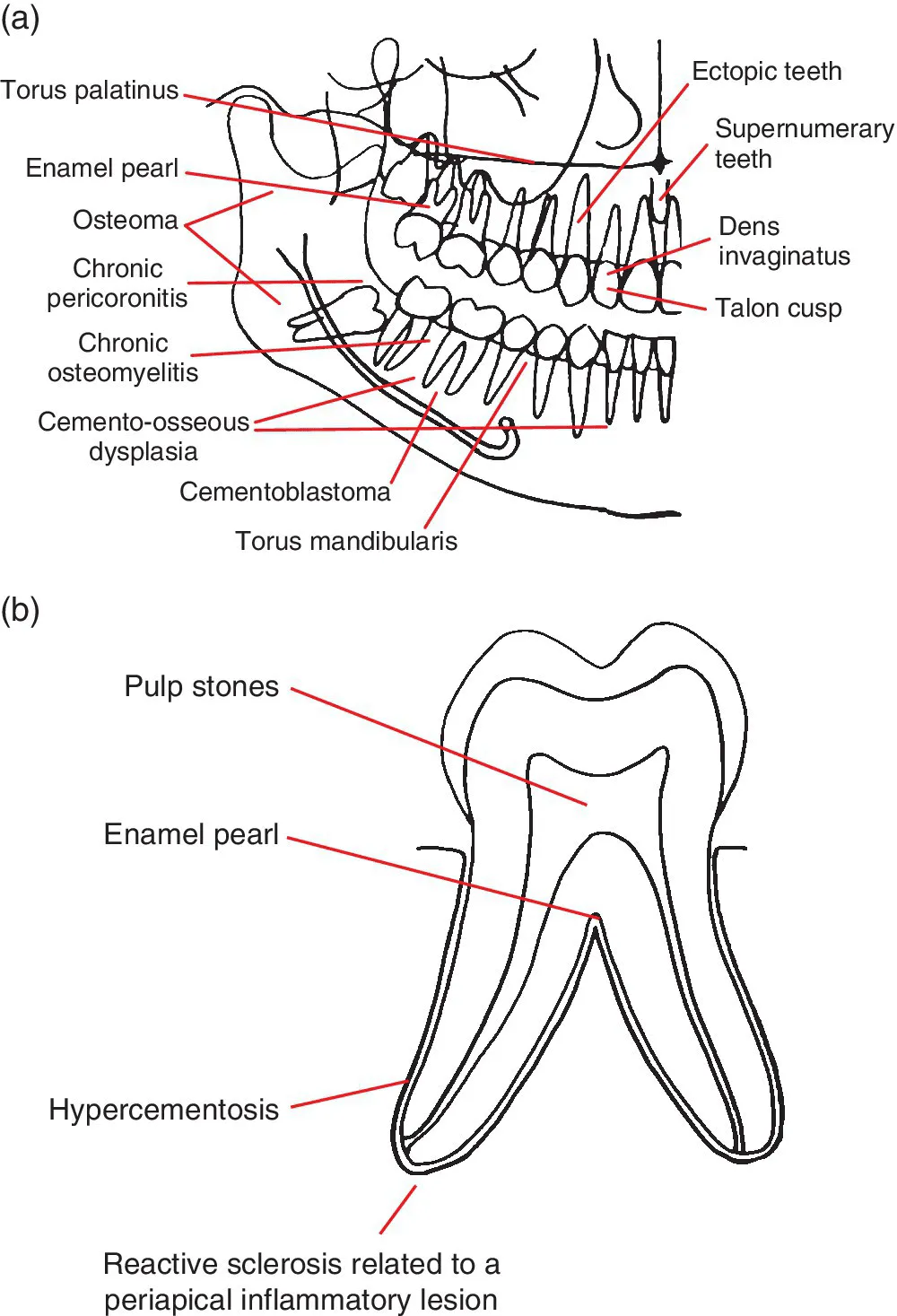
Figure 1.1 (a) Representation of the jaws and teeth and (b) larger representation of the fully erupted tooth. Conditions that have a predilection for certain regions of the jaws and teeth are shown. Note: (1) These lesions are not necessarily more common than other conditions. See the text for lists of common and less common conditions. (2) Most of these lesions also occur elsewhere within the jaws. (3) The pointers identify a region, not a specific site.
1.2 Lucent lesions of the jaws
For conditions affecting the TMJ, nasal cavity, paranasal sinuses, upper airway morphology, skull base and cervical spine, please refer to the dedicated chapters.
Common conditions
- Caries (see section 4.1)
- Periodontal bone loss (see section 5....
Inhaltsverzeichnis
- Cover
- Title Page
- Table of Contents
- List of Contributors
- Preface
- Acknowledgements
- How to Use This Atlas
- CHAPTER 1: Problem Solving Diagrams
- CHAPTER 2: Radiological Anatomy
- CHAPTER 3: Anomalies Related to the Teeth
- CHAPTER 4: Conditions Related to Loss of Tooth Structure
- CHAPTER 5: Inflammatory Lesions of the Jaws
- CHAPTER 6: Osteoradionecrosis and Osteonecrosis of the Jaws
- CHAPTER 7: Hamartomatous/Hyperplastic Bony Opacities and Prominences Involving the Jaws
- CHAPTER 8: Cysts and Cyst‐like Lesions Involving the Jaws
- CHAPTER 9: Fibro‐osseous Lesions of the Jaws
- CHAPTER 10: Benign Tumours Involving the Jaws
- CHAPTER 11: Malignant Tumours Involving the Jaws
- CHAPTER 12: Vascular Anomalies of the Mid‐ and Lower Face
- CHAPTER 13: Other Diseases Affecting the Jaws
- CHAPTER 14: Other Morphological Anomalies Involving the Jaws
- CHAPTER 15: Other Systemic Disorders that may Involve the Jaws
- CHAPTER 16: Common Opacities in the Orofacial Soft Tissues
- CHAPTER 17: Trauma and Fractures
- CHAPTER 18: Temporomandibular Joints
- CHAPTER 19: Nasal Cavity, Paranasal Sinuses and Upper Aerodigestive Tract Impressions
- CHAPTER 20: The Skull Base
- CHAPTER 21: The Cervical Spine
- Index
- End User License Agreement
Zitierstile für Atlas of Oral and Maxillofacial Radiology
APA 6 Citation
Koong, B. (2017). Atlas of Oral and Maxillofacial Radiology (1st ed.). Wiley. Retrieved from https://www.perlego.com/book/991897/atlas-of-oral-and-maxillofacial-radiology-pdf (Original work published 2017)
Chicago Citation
Koong, Bernard. (2017) 2017. Atlas of Oral and Maxillofacial Radiology. 1st ed. Wiley. https://www.perlego.com/book/991897/atlas-of-oral-and-maxillofacial-radiology-pdf.
Harvard Citation
Koong, B. (2017) Atlas of Oral and Maxillofacial Radiology. 1st edn. Wiley. Available at: https://www.perlego.com/book/991897/atlas-of-oral-and-maxillofacial-radiology-pdf (Accessed: 14 October 2022).
MLA 7 Citation
Koong, Bernard. Atlas of Oral and Maxillofacial Radiology. 1st ed. Wiley, 2017. Web. 14 Oct. 2022.