eBook - ePub
Atlas of Oral and Maxillofacial Radiology
Bernard Koong
This is a test
- English
- ePUB (apto para móviles)
- Disponible en iOS y Android
eBook - ePub
Atlas of Oral and Maxillofacial Radiology
Bernard Koong
Detalles del libro
Vista previa del libro
Índice
Citas
Información del libro
The Atlas of Oral and Maxillofacial Radiology presents an extensive case collection of both common and less common conditions of the jaws and teeth. Focusing on the essentials of radiologic interpretation, this is a go-to companion for clinicians in everyday practice who have radiologically identified a potential abnormality, as well as a comprehensive study guide for students at all levels of dentistry, surgery and radiology.
- Unique lesion-based problem solving chapter makes this an easy-to-use reference in a clinical setting
- Includes 2D intraoral radiography, the panoramic radiograph, cone beam CT, multidetector CT and MRI
- Multiple cases are presented in order to demonstrate the variation in the radiological appearances of conditions affecting the jaws and teeth
- Special focus on conditions where diagnostic imaging may substantially contribute to diagnosis
- Features a useful chapter covering the temporomandibular joint
Preguntas frecuentes
¿Cómo cancelo mi suscripción?
¿Cómo descargo los libros?
Por el momento, todos nuestros libros ePub adaptables a dispositivos móviles se pueden descargar a través de la aplicación. La mayor parte de nuestros PDF también se puede descargar y ya estamos trabajando para que el resto también sea descargable. Obtén más información aquí.
¿En qué se diferencian los planes de precios?
Ambos planes te permiten acceder por completo a la biblioteca y a todas las funciones de Perlego. Las únicas diferencias son el precio y el período de suscripción: con el plan anual ahorrarás en torno a un 30 % en comparación con 12 meses de un plan mensual.
¿Qué es Perlego?
Somos un servicio de suscripción de libros de texto en línea que te permite acceder a toda una biblioteca en línea por menos de lo que cuesta un libro al mes. Con más de un millón de libros sobre más de 1000 categorías, ¡tenemos todo lo que necesitas! Obtén más información aquí.
¿Perlego ofrece la función de texto a voz?
Busca el símbolo de lectura en voz alta en tu próximo libro para ver si puedes escucharlo. La herramienta de lectura en voz alta lee el texto en voz alta por ti, resaltando el texto a medida que se lee. Puedes pausarla, acelerarla y ralentizarla. Obtén más información aquí.
¿Es Atlas of Oral and Maxillofacial Radiology un PDF/ePUB en línea?
Sí, puedes acceder a Atlas of Oral and Maxillofacial Radiology de Bernard Koong en formato PDF o ePUB, así como a otros libros populares de Medicine y Dentistry. Tenemos más de un millón de libros disponibles en nuestro catálogo para que explores.
Información
CHAPTER 1
Problem Solving Diagrams
1.1 Opaque and largely opaque conditions related to the jaws
For conditions affecting the temporomandibular joint (TMJ), nasal cavity, paranasal sinuses, upper airway morphology, skull base and cervical spine, please refer to the dedicated chapters.
On plain films, including panoramic and cephalometric radiographs, soft tissue calcifications may be projected over the jaws (see Chapter 16).
Common conditions
- Reactive sclerosis related to a periapical inflammatory lesion (see section 5.1)
- Bone island (see section 7.4)
- Exostoses (see section 7.3)
- Torus palatinus (see section 7.1)
- Torus mandibularis (see section 7.2)
- Ectopic teeth (see section 3.4)
- Chronic pericoronitis (see section 5.3)
- Supernumerary teeth (see section 3.1)
- Cemento‐osseous dysplasia including periapical osseous dysplasia (see section 9.2)
- Pulp stones (see section 3.21)
- Hypercementosis (see section 3.22)
- Odontoma (see section 10.3)
- Dens invaginatus (see section 3.11)
- Fibrous dysplasia (see section 9.1)
- Enamel pearl (see section 3.9)
- Talon cusp (see section 3.10)
Less common conditions
- Osteoma (see section 10.10)
- Malignant lesions including metastatic disease (see sections 11.1–11.3)
- Chronic osteomyelitis (see section 5.4)
- Ossifying fibroma (see section 9.3)
- Cementoblastoma (see section 10.9)
- Osteoblastoma (see section 10.14)
- Osteoid osteoma (see section 10.15)
- Paget disease of bone (see section 13.5)
- Osteopetrosis (see section 15.2)
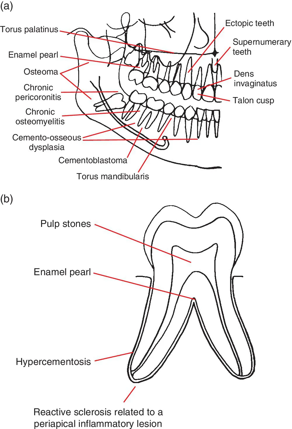
Figure 1.1 (a) Representation of the jaws and teeth and (b) larger representation of the fully erupted tooth. Conditions that have a predilection for certain regions of the jaws and teeth are shown. Note: (1) These lesions are not necessarily more common than other conditions. See the text for lists of common and less common conditions. (2) Most of these lesions also occur elsewhere within the jaws. (3) The pointers identify a region, not a specific site.
1.2 Lucent lesions of the jaws
For conditions affecting the TMJ, nasal cavity, paranasal sinuses, upper airway morphology, skull base and cervical spine, please refer to the dedicated chapters.
Common conditions
- Caries (see section 4.1)
- Periodontal bone loss (see section 5....
Índice
- Cover
- Title Page
- Table of Contents
- List of Contributors
- Preface
- Acknowledgements
- How to Use This Atlas
- CHAPTER 1: Problem Solving Diagrams
- CHAPTER 2: Radiological Anatomy
- CHAPTER 3: Anomalies Related to the Teeth
- CHAPTER 4: Conditions Related to Loss of Tooth Structure
- CHAPTER 5: Inflammatory Lesions of the Jaws
- CHAPTER 6: Osteoradionecrosis and Osteonecrosis of the Jaws
- CHAPTER 7: Hamartomatous/Hyperplastic Bony Opacities and Prominences Involving the Jaws
- CHAPTER 8: Cysts and Cyst‐like Lesions Involving the Jaws
- CHAPTER 9: Fibro‐osseous Lesions of the Jaws
- CHAPTER 10: Benign Tumours Involving the Jaws
- CHAPTER 11: Malignant Tumours Involving the Jaws
- CHAPTER 12: Vascular Anomalies of the Mid‐ and Lower Face
- CHAPTER 13: Other Diseases Affecting the Jaws
- CHAPTER 14: Other Morphological Anomalies Involving the Jaws
- CHAPTER 15: Other Systemic Disorders that may Involve the Jaws
- CHAPTER 16: Common Opacities in the Orofacial Soft Tissues
- CHAPTER 17: Trauma and Fractures
- CHAPTER 18: Temporomandibular Joints
- CHAPTER 19: Nasal Cavity, Paranasal Sinuses and Upper Aerodigestive Tract Impressions
- CHAPTER 20: The Skull Base
- CHAPTER 21: The Cervical Spine
- Index
- End User License Agreement
Estilos de citas para Atlas of Oral and Maxillofacial Radiology
APA 6 Citation
Koong, B. (2017). Atlas of Oral and Maxillofacial Radiology (1st ed.). Wiley. Retrieved from https://www.perlego.com/book/991897/atlas-of-oral-and-maxillofacial-radiology-pdf (Original work published 2017)
Chicago Citation
Koong, Bernard. (2017) 2017. Atlas of Oral and Maxillofacial Radiology. 1st ed. Wiley. https://www.perlego.com/book/991897/atlas-of-oral-and-maxillofacial-radiology-pdf.
Harvard Citation
Koong, B. (2017) Atlas of Oral and Maxillofacial Radiology. 1st edn. Wiley. Available at: https://www.perlego.com/book/991897/atlas-of-oral-and-maxillofacial-radiology-pdf (Accessed: 14 October 2022).
MLA 7 Citation
Koong, Bernard. Atlas of Oral and Maxillofacial Radiology. 1st ed. Wiley, 2017. Web. 14 Oct. 2022.