eBook - ePub
Atlas of Oral and Maxillofacial Radiology
Bernard Koong
This is a test
- English
- ePUB (adapté aux mobiles)
- Disponible sur iOS et Android
eBook - ePub
Atlas of Oral and Maxillofacial Radiology
Bernard Koong
Détails du livre
Aperçu du livre
Table des matières
Citations
À propos de ce livre
The Atlas of Oral and Maxillofacial Radiology presents an extensive case collection of both common and less common conditions of the jaws and teeth. Focusing on the essentials of radiologic interpretation, this is a go-to companion for clinicians in everyday practice who have radiologically identified a potential abnormality, as well as a comprehensive study guide for students at all levels of dentistry, surgery and radiology.
- Unique lesion-based problem solving chapter makes this an easy-to-use reference in a clinical setting
- Includes 2D intraoral radiography, the panoramic radiograph, cone beam CT, multidetector CT and MRI
- Multiple cases are presented in order to demonstrate the variation in the radiological appearances of conditions affecting the jaws and teeth
- Special focus on conditions where diagnostic imaging may substantially contribute to diagnosis
- Features a useful chapter covering the temporomandibular joint
Foire aux questions
Comment puis-je résilier mon abonnement ?
Il vous suffit de vous rendre dans la section compte dans paramètres et de cliquer sur « Résilier l’abonnement ». C’est aussi simple que cela ! Une fois que vous aurez résilié votre abonnement, il restera actif pour le reste de la période pour laquelle vous avez payé. Découvrez-en plus ici.
Puis-je / comment puis-je télécharger des livres ?
Pour le moment, tous nos livres en format ePub adaptés aux mobiles peuvent être téléchargés via l’application. La plupart de nos PDF sont également disponibles en téléchargement et les autres seront téléchargeables très prochainement. Découvrez-en plus ici.
Quelle est la différence entre les formules tarifaires ?
Les deux abonnements vous donnent un accès complet à la bibliothèque et à toutes les fonctionnalités de Perlego. Les seules différences sont les tarifs ainsi que la période d’abonnement : avec l’abonnement annuel, vous économiserez environ 30 % par rapport à 12 mois d’abonnement mensuel.
Qu’est-ce que Perlego ?
Nous sommes un service d’abonnement à des ouvrages universitaires en ligne, où vous pouvez accéder à toute une bibliothèque pour un prix inférieur à celui d’un seul livre par mois. Avec plus d’un million de livres sur plus de 1 000 sujets, nous avons ce qu’il vous faut ! Découvrez-en plus ici.
Prenez-vous en charge la synthèse vocale ?
Recherchez le symbole Écouter sur votre prochain livre pour voir si vous pouvez l’écouter. L’outil Écouter lit le texte à haute voix pour vous, en surlignant le passage qui est en cours de lecture. Vous pouvez le mettre sur pause, l’accélérer ou le ralentir. Découvrez-en plus ici.
Est-ce que Atlas of Oral and Maxillofacial Radiology est un PDF/ePUB en ligne ?
Oui, vous pouvez accéder à Atlas of Oral and Maxillofacial Radiology par Bernard Koong en format PDF et/ou ePUB ainsi qu’à d’autres livres populaires dans Medicine et Dentistry. Nous disposons de plus d’un million d’ouvrages à découvrir dans notre catalogue.
Informations
CHAPTER 1
Problem Solving Diagrams
1.1 Opaque and largely opaque conditions related to the jaws
For conditions affecting the temporomandibular joint (TMJ), nasal cavity, paranasal sinuses, upper airway morphology, skull base and cervical spine, please refer to the dedicated chapters.
On plain films, including panoramic and cephalometric radiographs, soft tissue calcifications may be projected over the jaws (see Chapter 16).
Common conditions
- Reactive sclerosis related to a periapical inflammatory lesion (see section 5.1)
- Bone island (see section 7.4)
- Exostoses (see section 7.3)
- Torus palatinus (see section 7.1)
- Torus mandibularis (see section 7.2)
- Ectopic teeth (see section 3.4)
- Chronic pericoronitis (see section 5.3)
- Supernumerary teeth (see section 3.1)
- Cemento‐osseous dysplasia including periapical osseous dysplasia (see section 9.2)
- Pulp stones (see section 3.21)
- Hypercementosis (see section 3.22)
- Odontoma (see section 10.3)
- Dens invaginatus (see section 3.11)
- Fibrous dysplasia (see section 9.1)
- Enamel pearl (see section 3.9)
- Talon cusp (see section 3.10)
Less common conditions
- Osteoma (see section 10.10)
- Malignant lesions including metastatic disease (see sections 11.1–11.3)
- Chronic osteomyelitis (see section 5.4)
- Ossifying fibroma (see section 9.3)
- Cementoblastoma (see section 10.9)
- Osteoblastoma (see section 10.14)
- Osteoid osteoma (see section 10.15)
- Paget disease of bone (see section 13.5)
- Osteopetrosis (see section 15.2)
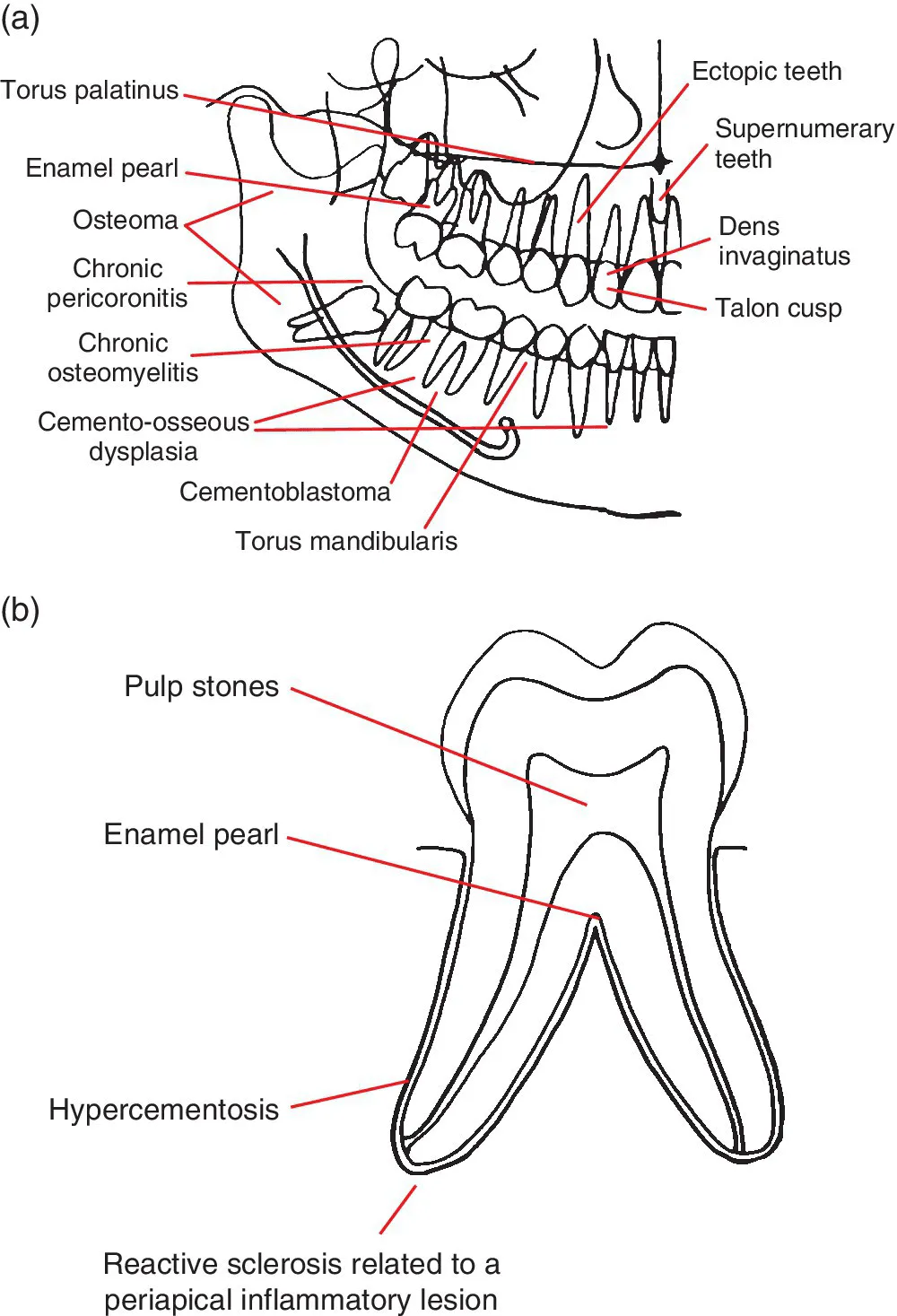
Figure 1.1 (a) Representation of the jaws and teeth and (b) larger representation of the fully erupted tooth. Conditions that have a predilection for certain regions of the jaws and teeth are shown. Note: (1) These lesions are not necessarily more common than other conditions. See the text for lists of common and less common conditions. (2) Most of these lesions also occur elsewhere within the jaws. (3) The pointers identify a region, not a specific site.
1.2 Lucent lesions of the jaws
For conditions affecting the TMJ, nasal cavity, paranasal sinuses, upper airway morphology, skull base and cervical spine, please refer to the dedicated chapters.
Common conditions
- Caries (see section 4.1)
- Periodontal bone loss (see section 5....
Table des matières
- Cover
- Title Page
- Table of Contents
- List of Contributors
- Preface
- Acknowledgements
- How to Use This Atlas
- CHAPTER 1: Problem Solving Diagrams
- CHAPTER 2: Radiological Anatomy
- CHAPTER 3: Anomalies Related to the Teeth
- CHAPTER 4: Conditions Related to Loss of Tooth Structure
- CHAPTER 5: Inflammatory Lesions of the Jaws
- CHAPTER 6: Osteoradionecrosis and Osteonecrosis of the Jaws
- CHAPTER 7: Hamartomatous/Hyperplastic Bony Opacities and Prominences Involving the Jaws
- CHAPTER 8: Cysts and Cyst‐like Lesions Involving the Jaws
- CHAPTER 9: Fibro‐osseous Lesions of the Jaws
- CHAPTER 10: Benign Tumours Involving the Jaws
- CHAPTER 11: Malignant Tumours Involving the Jaws
- CHAPTER 12: Vascular Anomalies of the Mid‐ and Lower Face
- CHAPTER 13: Other Diseases Affecting the Jaws
- CHAPTER 14: Other Morphological Anomalies Involving the Jaws
- CHAPTER 15: Other Systemic Disorders that may Involve the Jaws
- CHAPTER 16: Common Opacities in the Orofacial Soft Tissues
- CHAPTER 17: Trauma and Fractures
- CHAPTER 18: Temporomandibular Joints
- CHAPTER 19: Nasal Cavity, Paranasal Sinuses and Upper Aerodigestive Tract Impressions
- CHAPTER 20: The Skull Base
- CHAPTER 21: The Cervical Spine
- Index
- End User License Agreement
Normes de citation pour Atlas of Oral and Maxillofacial Radiology
APA 6 Citation
Koong, B. (2017). Atlas of Oral and Maxillofacial Radiology (1st ed.). Wiley. Retrieved from https://www.perlego.com/book/991897/atlas-of-oral-and-maxillofacial-radiology-pdf (Original work published 2017)
Chicago Citation
Koong, Bernard. (2017) 2017. Atlas of Oral and Maxillofacial Radiology. 1st ed. Wiley. https://www.perlego.com/book/991897/atlas-of-oral-and-maxillofacial-radiology-pdf.
Harvard Citation
Koong, B. (2017) Atlas of Oral and Maxillofacial Radiology. 1st edn. Wiley. Available at: https://www.perlego.com/book/991897/atlas-of-oral-and-maxillofacial-radiology-pdf (Accessed: 14 October 2022).
MLA 7 Citation
Koong, Bernard. Atlas of Oral and Maxillofacial Radiology. 1st ed. Wiley, 2017. Web. 14 Oct. 2022.