eBook - ePub
Atlas of Oral and Maxillofacial Radiology
Bernard Koong
This is a test
- English
- ePUB (disponibile sull'app)
- Disponibile su iOS e Android
eBook - ePub
Atlas of Oral and Maxillofacial Radiology
Bernard Koong
Dettagli del libro
Anteprima del libro
Indice dei contenuti
Citazioni
Informazioni sul libro
The Atlas of Oral and Maxillofacial Radiology presents an extensive case collection of both common and less common conditions of the jaws and teeth. Focusing on the essentials of radiologic interpretation, this is a go-to companion for clinicians in everyday practice who have radiologically identified a potential abnormality, as well as a comprehensive study guide for students at all levels of dentistry, surgery and radiology.
- Unique lesion-based problem solving chapter makes this an easy-to-use reference in a clinical setting
- Includes 2D intraoral radiography, the panoramic radiograph, cone beam CT, multidetector CT and MRI
- Multiple cases are presented in order to demonstrate the variation in the radiological appearances of conditions affecting the jaws and teeth
- Special focus on conditions where diagnostic imaging may substantially contribute to diagnosis
- Features a useful chapter covering the temporomandibular joint
Domande frequenti
Come faccio ad annullare l'abbonamento?
È semplicissimo: basta accedere alla sezione Account nelle Impostazioni e cliccare su "Annulla abbonamento". Dopo la cancellazione, l'abbonamento rimarrà attivo per il periodo rimanente già pagato. Per maggiori informazioni, clicca qui
È possibile scaricare libri? Se sì, come?
Al momento è possibile scaricare tramite l'app tutti i nostri libri ePub mobile-friendly. Anche la maggior parte dei nostri PDF è scaricabile e stiamo lavorando per rendere disponibile quanto prima il download di tutti gli altri file. Per maggiori informazioni, clicca qui
Che differenza c'è tra i piani?
Entrambi i piani ti danno accesso illimitato alla libreria e a tutte le funzionalità di Perlego. Le uniche differenze sono il prezzo e il periodo di abbonamento: con il piano annuale risparmierai circa il 30% rispetto a 12 rate con quello mensile.
Cos'è Perlego?
Perlego è un servizio di abbonamento a testi accademici, che ti permette di accedere a un'intera libreria online a un prezzo inferiore rispetto a quello che pagheresti per acquistare un singolo libro al mese. Con oltre 1 milione di testi suddivisi in più di 1.000 categorie, troverai sicuramente ciò che fa per te! Per maggiori informazioni, clicca qui.
Perlego supporta la sintesi vocale?
Cerca l'icona Sintesi vocale nel prossimo libro che leggerai per verificare se è possibile riprodurre l'audio. Questo strumento permette di leggere il testo a voce alta, evidenziandolo man mano che la lettura procede. Puoi aumentare o diminuire la velocità della sintesi vocale, oppure sospendere la riproduzione. Per maggiori informazioni, clicca qui.
Atlas of Oral and Maxillofacial Radiology è disponibile online in formato PDF/ePub?
Sì, puoi accedere a Atlas of Oral and Maxillofacial Radiology di Bernard Koong in formato PDF e/o ePub, così come ad altri libri molto apprezzati nelle sezioni relative a Medicine e Dentistry. Scopri oltre 1 milione di libri disponibili nel nostro catalogo.
Informazioni
CHAPTER 1
Problem Solving Diagrams
1.1 Opaque and largely opaque conditions related to the jaws
For conditions affecting the temporomandibular joint (TMJ), nasal cavity, paranasal sinuses, upper airway morphology, skull base and cervical spine, please refer to the dedicated chapters.
On plain films, including panoramic and cephalometric radiographs, soft tissue calcifications may be projected over the jaws (see Chapter 16).
Common conditions
- Reactive sclerosis related to a periapical inflammatory lesion (see section 5.1)
- Bone island (see section 7.4)
- Exostoses (see section 7.3)
- Torus palatinus (see section 7.1)
- Torus mandibularis (see section 7.2)
- Ectopic teeth (see section 3.4)
- Chronic pericoronitis (see section 5.3)
- Supernumerary teeth (see section 3.1)
- Cemento‐osseous dysplasia including periapical osseous dysplasia (see section 9.2)
- Pulp stones (see section 3.21)
- Hypercementosis (see section 3.22)
- Odontoma (see section 10.3)
- Dens invaginatus (see section 3.11)
- Fibrous dysplasia (see section 9.1)
- Enamel pearl (see section 3.9)
- Talon cusp (see section 3.10)
Less common conditions
- Osteoma (see section 10.10)
- Malignant lesions including metastatic disease (see sections 11.1–11.3)
- Chronic osteomyelitis (see section 5.4)
- Ossifying fibroma (see section 9.3)
- Cementoblastoma (see section 10.9)
- Osteoblastoma (see section 10.14)
- Osteoid osteoma (see section 10.15)
- Paget disease of bone (see section 13.5)
- Osteopetrosis (see section 15.2)
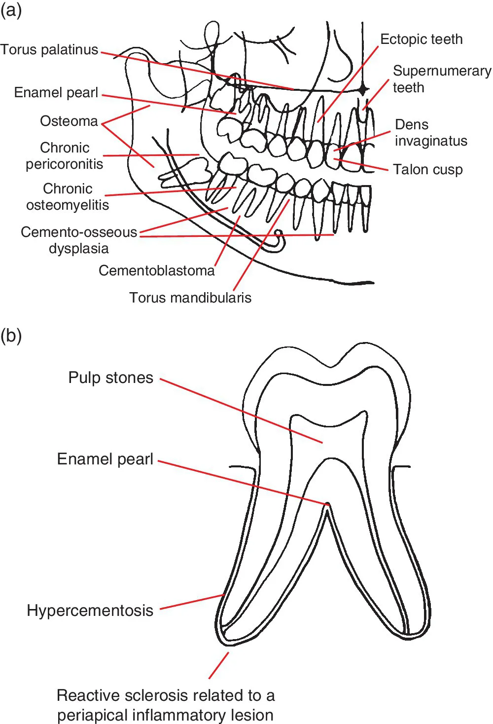
Figure 1.1 (a) Representation of the jaws and teeth and (b) larger representation of the fully erupted tooth. Conditions that have a predilection for certain regions of the jaws and teeth are shown. Note: (1) These lesions are not necessarily more common than other conditions. See the text for lists of common and less common conditions. (2) Most of these lesions also occur elsewhere within the jaws. (3) The pointers identify a region, not a specific site.
1.2 Lucent lesions of the jaws
For conditions affecting the TMJ, nasal cavity, paranasal sinuses, upper airway morphology, skull base and cervical spine, please refer to the dedicated chapters.
Common conditions
- Caries (see section 4.1)
- Periodontal bone loss (see section 5....
Indice dei contenuti
- Cover
- Title Page
- Table of Contents
- List of Contributors
- Preface
- Acknowledgements
- How to Use This Atlas
- CHAPTER 1: Problem Solving Diagrams
- CHAPTER 2: Radiological Anatomy
- CHAPTER 3: Anomalies Related to the Teeth
- CHAPTER 4: Conditions Related to Loss of Tooth Structure
- CHAPTER 5: Inflammatory Lesions of the Jaws
- CHAPTER 6: Osteoradionecrosis and Osteonecrosis of the Jaws
- CHAPTER 7: Hamartomatous/Hyperplastic Bony Opacities and Prominences Involving the Jaws
- CHAPTER 8: Cysts and Cyst‐like Lesions Involving the Jaws
- CHAPTER 9: Fibro‐osseous Lesions of the Jaws
- CHAPTER 10: Benign Tumours Involving the Jaws
- CHAPTER 11: Malignant Tumours Involving the Jaws
- CHAPTER 12: Vascular Anomalies of the Mid‐ and Lower Face
- CHAPTER 13: Other Diseases Affecting the Jaws
- CHAPTER 14: Other Morphological Anomalies Involving the Jaws
- CHAPTER 15: Other Systemic Disorders that may Involve the Jaws
- CHAPTER 16: Common Opacities in the Orofacial Soft Tissues
- CHAPTER 17: Trauma and Fractures
- CHAPTER 18: Temporomandibular Joints
- CHAPTER 19: Nasal Cavity, Paranasal Sinuses and Upper Aerodigestive Tract Impressions
- CHAPTER 20: The Skull Base
- CHAPTER 21: The Cervical Spine
- Index
- End User License Agreement
Stili delle citazioni per Atlas of Oral and Maxillofacial Radiology
APA 6 Citation
Koong, B. (2017). Atlas of Oral and Maxillofacial Radiology (1st ed.). Wiley. Retrieved from https://www.perlego.com/book/991897/atlas-of-oral-and-maxillofacial-radiology-pdf (Original work published 2017)
Chicago Citation
Koong, Bernard. (2017) 2017. Atlas of Oral and Maxillofacial Radiology. 1st ed. Wiley. https://www.perlego.com/book/991897/atlas-of-oral-and-maxillofacial-radiology-pdf.
Harvard Citation
Koong, B. (2017) Atlas of Oral and Maxillofacial Radiology. 1st edn. Wiley. Available at: https://www.perlego.com/book/991897/atlas-of-oral-and-maxillofacial-radiology-pdf (Accessed: 14 October 2022).
MLA 7 Citation
Koong, Bernard. Atlas of Oral and Maxillofacial Radiology. 1st ed. Wiley, 2017. Web. 14 Oct. 2022.